Author: MSc Marcin Goras – Master of Public Health, Specialization in Emergency Medical Services
Published: September 16, 2025
Last Updated: September 16, 2025
Reading Time: 12-15 minutes
Introduction
Ventricular tachycardia (VT) represents one of the most serious cardiac rhythm disorders, potentially threatening life within minutes if left untreated. Medical literature indicates that this condition affects thousands of individuals worldwide, ranging from those with underlying heart disease to apparently healthy individuals with inherited conditions. Understanding ventricular tachycardia is crucial for recognizing warning signs and seeking immediate medical attention when necessary.
Contemporary cardiology has made remarkable advances in both understanding and treating ventricular tachycardia. Studies demonstrate that early recognition, proper risk stratification, and appropriate therapeutic interventions can significantly improve patient outcomes and quality of life. This comprehensive guide provides evidence-based information while emphasizing the critical importance of professional medical evaluation and treatment planning.
Research shows that ventricular tachycardia can present in various forms, from brief, self-terminating episodes to sustained arrhythmias requiring emergency intervention. The condition’s complexity necessitates individualized assessment and treatment approaches based on multiple clinical factors.
What is Ventricular Tachycardia?
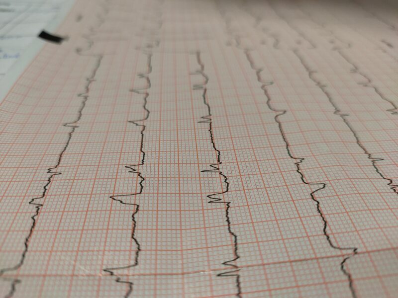
Ventricular tachycardia is a rapid heart rhythm originating from the ventricles, the heart’s main pumping chambers. Clinical studies define VT as three or more consecutive ventricular beats occurring at rates typically exceeding 100 beats per minute, often reaching 150-250 beats per minute. Research indicates that this abnormal rhythm can significantly compromise the heart’s ability to pump blood effectively.
Key Characteristics of VT
| Parameter | Normal Heart Rhythm | Ventricular Tachycardia |
|---|---|---|
| Heart Rate | 60-100 bpm | 150-250 bpm |
| Origin | Atrial (SA node) | Ventricular tissue |
| QRS Duration | <120 milliseconds | >120 milliseconds |
| Hemodynamic Impact | Stable | Often compromised |
| Duration Significance | N/A | <30 sec (NSVT) vs >30 sec (SVT) |
Medical literature emphasizes that ventricular tachycardia differs fundamentally from other rapid heart rhythms because it originates below the atrioventricular (AV) node, directly from ventricular muscle or specialized conduction tissue. This origin significantly impacts both the electrocardiographic appearance and clinical consequences.
Clinical Significance
Studies demonstrate that ventricular tachycardia can rapidly progress to ventricular fibrillation, a chaotic rhythm incompatible with life. Research indicates that the hemodynamic consequences depend on multiple factors including the heart rate, underlying cardiac function, and the presence of structural heart disease.
Understanding Heart Electrical System
Comprehensive understanding of ventricular tachycardia requires knowledge of the heart’s normal electrical conduction system. Research shows that the heart’s electrical activity normally begins in the sinoatrial (SA) node, located in the right atrium, which serves as the natural pacemaker.
Normal Electrical Conduction Pathway
- Sinoatrial Node: Initiates electrical impulses at 60-100 beats per minute
- Atrial Conduction: Impulses spread through both atria
- Atrioventricular Node: Provides brief delay allowing ventricular filling
- Bundle of His: Conducts impulses to ventricular conduction system
- Bundle Branches: Right and left branches distribute impulses
- Purkinje Fibers: Fine network ensuring coordinated ventricular contraction
VT Development Mechanisms
Medical literature identifies several mechanisms by which ventricular tachycardia can develop:
Reentry Circuits: Studies show this is the most common mechanism, where electrical impulses circulate repeatedly through abnormal pathways, often around areas of cardiac scar tissue.
Abnormal Automaticity: Research indicates that certain ventricular cells can develop abnormal pacemaker activity, firing rapidly and overriding normal rhythm.
Triggered Activity: Evidence suggests that abnormal calcium handling within cardiac cells can trigger rapid, repetitive firing.
Types and Classifications of VT
Contemporary cardiology classifies ventricular tachycardia based on several important characteristics that influence treatment decisions and prognosis.
Duration-Based Classification
Non-Sustained Ventricular Tachycardia (NSVT)
- Duration: Less than 30 seconds
- Clinical significance: Studies suggest variable risk depending on underlying heart disease
- Symptoms: Often asymptomatic or minimal palpitations
Sustained Ventricular Tachycardia (SVT)
- Duration: 30 seconds or longer, or requiring intervention
- Clinical significance: Research indicates higher risk of hemodynamic compromise
- Symptoms: Often symptomatic with potential for rapid deterioration
Morphological Classification
Monomorphic VT
- Characteristics: Consistent QRS morphology throughout the episode
- Mechanism: Typically reentrant, often related to cardiac scar
- Clinical context: Studies show strong association with structural heart disease
Polymorphic VT
- Characteristics: Changing QRS morphology during the episode
- Subtypes: Includes torsades de pointes when associated with QT prolongation
- Clinical context: Research indicates association with acute ischemia or channelopathies
Hemodynamic Classification
Medical literature emphasizes the importance of hemodynamic assessment:
Hemodynamically Stable VT: Patient maintains adequate blood pressure and consciousness Hemodynamically Unstable VT: Associated with hypotension, altered consciousness, or cardiac arrest
Underlying Causes and Risk Factors
Research identifies numerous conditions and factors that can predispose individuals to ventricular tachycardia development.
Structural Heart Disease
Coronary Artery Disease
- Studies indicate this is the most common underlying cause
- Mechanism: Scar tissue from previous myocardial infarction creates reentry circuits
- Risk factors: Prior heart attack significantly increases VT risk
Cardiomyopathies
- Ischemic cardiomyopathy: Research shows high VT risk in patients with reduced ejection fraction
- Dilated cardiomyopathy: Studies demonstrate significant arrhythmia risk
- Hypertrophic cardiomyopathy: Evidence indicates risk varies by phenotype
- Arrhythmogenic cardiomyopathy: Research shows particularly high VT risk
Valvular Heart Disease
- Aortic stenosis and mitral regurgitation can predispose to VT
- Studies suggest risk increases with severity and duration
Inherited Conditions
Channelopathies
- Long QT syndrome: Research indicates risk of polymorphic VT/torsades de pointes
- Brugada syndrome: Studies show risk of rapid VT/VF
- Catecholaminergic polymorphic VT: Evidence demonstrates exercise-induced VT risk
Acquired Conditions
Electrolyte Imbalances
- Hypokalemia, hypomagnesemia, and hypocalcemia
- Studies emphasize the importance of electrolyte monitoring
Medication-Related
- Antiarrhythmic drugs (paradoxically)
- QT-prolonging medications
- Research indicates numerous drugs can trigger VT
Metabolic Disorders
- Thyroid dysfunction
- Diabetes mellitus complications
- Studies suggest multiple metabolic pathways can influence VT risk
Acute Triggers
Medical literature identifies several acute factors that can precipitate VT:
- Acute myocardial ischemia
- Electrolyte shifts
- Sympathetic stimulation
- Hypoxia and acidosis
Clinical Symptoms and Warning Signs
Research demonstrates that ventricular tachycardia symptoms vary significantly based on the heart rate, duration, underlying cardiac function, and hemodynamic tolerance.
Common Symptoms
Cardiovascular Symptoms
- Palpitations: Studies indicate this is frequently reported, described as rapid, pounding heartbeat
- Chest pain: Research shows variable intensity, may indicate ischemia
- Shortness of breath: Evidence suggests correlation with reduced cardiac output
Neurological Symptoms
- Dizziness: Studies indicate common due to reduced cerebral perfusion
- Presyncope: Research shows warning sign of hemodynamic compromise
- Syncope: Evidence indicates serious symptom requiring immediate evaluation
Constitutional Symptoms
- Fatigue and weakness
- Exercise intolerance
- Studies suggest these may indicate chronic or recurrent episodes
Emergency Warning Signs
Medical literature emphasizes certain symptoms requiring immediate emergency care:
- Loss of consciousness
- Severe chest pain
- Severe shortness of breath
- Signs of cardiac arrest
- Rapid deterioration in clinical status
Asymptomatic Presentations
Research indicates that some patients, particularly those with preserved cardiac function, may experience minimal or no symptoms during brief VT episodes. However, studies emphasize that even asymptomatic VT can be clinically significant and require evaluation.
Diagnostic Methods
Contemporary cardiology employs multiple diagnostic approaches to identify, characterize, and risk-stratify patients with ventricular tachycardia.
Electrocardiographic Evaluation
12-Lead ECG
- Studies demonstrate this as the cornerstone of VT diagnosis
- Characteristics: Wide QRS complexes (>120 ms), rapid rate
- Research indicates specific morphology patterns can suggest VT origin
Diagnostic Criteria for VT
- AV dissociation (when visible)
- QRS width >140 ms in right bundle pattern, >160 ms in left bundle pattern
- Specific morphology criteria in various leads
- Studies emphasize importance of comparison with baseline ECG
Ambulatory Monitoring
Holter Monitoring
- 24-48 hour continuous recording
- Research shows useful for detecting asymptomatic episodes
- Studies indicate value in quantifying VT burden
Event Monitors
- Patient-activated or auto-triggered devices
- Evidence suggests useful for infrequent symptoms
- Research demonstrates improved diagnostic yield for episodic VT
Implantable Loop Recorders
- Long-term monitoring capability
- Studies show high diagnostic yield for infrequent episodes
- Research indicates valuable for syncope evaluation
Imaging Studies
Echocardiography
- Assessment of left ventricular function
- Detection of structural abnormalities
- Studies emphasize prognostic importance of ejection fraction
Cardiac MRI
- Detailed tissue characterization
- Scar detection and quantification
- Research demonstrates superior ability to identify arrhythmogenic substrate
Nuclear Imaging
- Perfusion and viability assessment
- Studies show value in ischemic cardiomyopathy evaluation
Invasive Testing
Electrophysiology Study
- Programmed ventricular stimulation
- Risk stratification capabilities
- Research indicates value for treatment planning
Coronary Angiography
- Assessment of coronary artery disease
- Studies emphasize importance in VT evaluation
Treatment Approaches
Medical management of ventricular tachycardia requires individualized approaches based on multiple factors including VT type, underlying disease, symptoms, and hemodynamic impact. It is essential to consult with qualified cardiologists or electrophysiologists for proper evaluation and treatment planning.
Acute Management Principles
Studies demonstrate that immediate treatment depends on hemodynamic stability:
Hemodynamically Stable VT
- Pharmacological cardioversion may be considered
- Continuous monitoring essential
- Research indicates importance of underlying cause treatment
Hemodynamically Unstable VT
- Immediate electrical cardioversion indicated
- Studies emphasize need for emergency intervention
- Advanced life support protocols apply
Pharmacological Approaches
Research indicates various therapeutic options exist, though specific treatments must be determined by healthcare providers:
Antiarrhythmic Medications
- Multiple drug classes available
- Studies suggest efficacy varies by VT mechanism and underlying disease
- Research emphasizes importance of individualized selection
Supportive Therapies
- Electrolyte replacement when indicated
- Beta-blocker therapy for appropriate candidates
- Studies show importance of heart failure optimization
Device-Based Therapies
Implantable Cardioverter Defibrillators (ICDs)
- Primary prevention in high-risk patients
- Secondary prevention after VT/VF survival
- Research demonstrates significant mortality benefit in appropriate candidates
Cardiac Resynchronization Therapy (CRT)
- For selected heart failure patients
- Studies suggest potential anti-arrhythmic benefits
- Research indicates improved outcomes when combined with ICD therapy
Catheter Ablation
Contemporary studies demonstrate that radiofrequency or cryoablation can effectively treat certain types of ventricular tachycardia:
Indications
- Recurrent VT despite medical therapy
- VT in patients with preserved cardiac function
- Research suggests growing role in VT management
Techniques
- Substrate mapping and modification
- Activation mapping for specific VT types
- Studies show evolving procedural approaches
Emergency Management
Medical literature emphasizes that ventricular tachycardia can represent a medical emergency requiring immediate intervention.
Recognition and Initial Assessment
Rapid Evaluation
- Assess hemodynamic stability
- Obtain 12-lead ECG when possible
- Studies emphasize importance of rapid decision-making
Hemodynamic Assessment
- Blood pressure measurement
- Level of consciousness evaluation
- Research indicates these factors guide immediate treatment
Treatment Algorithms
Stable VT Management
- Continuous cardiac monitoring
- Pharmacological cardioversion considerations
- Studies suggest importance of underlying cause identification
Unstable VT Management
- Immediate synchronized cardioversion
- Advanced cardiac life support protocols
- Research emphasizes need for rapid intervention
Post-Resuscitation Care
Medical literature indicates that survivors of VT episodes require:
- Comprehensive cardiac evaluation
- Risk stratification for recurrence
- Studies suggest importance of long-term management planning
Long-term Prognosis
Research demonstrates that the prognosis for patients with ventricular tachycardia varies significantly based on multiple factors.
Prognostic Factors
Favorable Indicators
- Normal or preserved cardiac function
- Absence of structural heart disease
- Successful treatment response
- Studies suggest better long-term outcomes
Concerning Factors
- Severely reduced ejection fraction
- Extensive structural heart disease
- Hemodynamically unstable presentations
- Research indicates need for aggressive management
Outcomes with Treatment
Contemporary studies suggest that with appropriate medical management:
- Many patients can achieve good symptomatic control
- Risk of sudden cardiac death can be significantly reduced
- Quality of life can be maintained or improved
- Research demonstrates importance of adherence to therapy
Monitoring Requirements
Medical literature emphasizes the need for:
- Regular cardiology follow-up
- Device monitoring when applicable
- Periodic risk reassessment
- Studies suggest evolving monitoring strategies
Prevention Strategies
Research identifies several approaches that may help reduce ventricular tachycardia risk:
Primary Prevention
Cardiovascular Risk Reduction
- Optimal management of coronary artery disease
- Blood pressure and cholesterol control
- Studies suggest importance of comprehensive cardiac care
Lifestyle Modifications
- Regular appropriate exercise
- Stress management techniques
- Research indicates multiple lifestyle factors influence cardiac health
Secondary Prevention
Medical Optimization
- Heart failure therapy when appropriate
- Electrolyte monitoring and replacement
- Studies emphasize importance of comprehensive medical management
Device Therapy
- ICD implantation for appropriate candidates
- Research demonstrates significant benefit for sudden death prevention
Risk Factor Management
Medical literature emphasizes:
- Treatment of underlying cardiac conditions
- Avoidance of known triggers
- Studies suggest importance of individualized approaches
Living with VT
Research demonstrates that many patients with ventricular tachycardia can maintain good quality of life with appropriate management.
Daily Life Considerations
Activity Modifications
- Exercise recommendations should be individualized
- Driving restrictions may apply
- Studies suggest importance of risk-based recommendations
Emergency Preparedness
- Family education about warning signs
- Emergency action plan development
- Research indicates importance of preparedness
Psychological Support
Medical literature recognizes that living with VT can be emotionally challenging:
- Anxiety about recurrent episodes
- Impact on family members
- Studies suggest benefit of psychological support
Follow-up Care
Research emphasizes the importance of:
- Regular medical monitoring
- Device follow-up when applicable
- Prompt reporting of symptoms
- Studies indicate evolving care models
FAQ
Q: Is ventricular tachycardia always dangerous? A: The clinical significance varies greatly. While some forms can be life-threatening emergencies, others may be better tolerated. Professional medical evaluation is essential to determine individual risk and appropriate management.
Q: Can VT be cured completely? A: Studies suggest that some types of VT can be successfully treated or eliminated with catheter ablation, while others require ongoing management. The approach depends on the underlying cause and VT characteristics.
Q: What should I do if I experience symptoms? A: Research emphasizes the importance of immediate medical evaluation for concerning symptoms, especially loss of consciousness, severe chest pain, or severe shortness of breath. Call emergency services for severe symptoms.
Q: Can exercise trigger VT? A: Studies indicate that exercise can trigger VT in some individuals, particularly those with certain inherited conditions. Exercise recommendations should be individualized based on medical evaluation.
Q: What is the difference between VT and other rapid heart rhythms? A: VT originates from the ventricles and typically produces wide QRS complexes on ECG, while other tachycardias usually originate above the ventricles. Professional evaluation is necessary for accurate diagnosis.
Q: How effective are ICDs in preventing sudden death? A: Research demonstrates that ICDs can significantly reduce the risk of sudden cardiac death in appropriate candidates. Studies show substantial mortality benefits in high-risk patients.
Q: Can medications prevent VT? A: Various medications can help prevent VT recurrence, though effectiveness depends on the specific type of VT and underlying conditions. Treatment decisions should always be made by qualified healthcare providers.
Medical Disclaimer
This article is provided for educational purposes only and does not constitute medical advice. The information presented should not be used for diagnosing or treating any medical condition. Ventricular tachycardia can be a medical emergency requiring immediate intervention. Individual cases vary significantly, and proper medical evaluation is essential for accurate diagnosis and appropriate treatment planning. Always consult with qualified healthcare providers for medical concerns, and seek immediate emergency medical attention for symptoms such as loss of consciousness, severe chest pain, or severe difficulty breathing. The author and publisher assume no responsibility for any consequences arising from the use of this information.
References
- Zeppenfeld K, Tfelt-Hansen J, de Riva M, et al. 2022 ESC Guidelines for the management of patients with ventricular arrhythmias and the prevention of sudden cardiac death. European Heart Journal. 2022;43(40):3997-4126.
- Al-Khatib SM, Stevenson WG, Ackerman MJ, et al. 2017 AHA/ACC/HRS Guideline for Management of Patients With Ventricular Arrhythmias and the Prevention of Sudden Cardiac Death. Circulation. 2018;138(13):e272-e391.
- Priori SG, Blomström-Lundqvist C, Mazzanti A, et al. 2015 ESC Guidelines for the management of patients with ventricular arrhythmias and the prevention of sudden cardiac death. European Heart Journal. 2015;36(41):2793-2867.
- Antzelevitch C, Burashnikov A. Overview of Basic Mechanisms of Cardiac Arrhythmia. Card Electrophysiol Rev. 2011;15(1):14-28.
- Tung R, Vaseghi M, Frankel DS, et al. Freedom from recurrent ventricular tachycardia after catheter ablation is associated with improved survival in patients with structural heart disease. Heart Rhythm. 2015;12(9):1997-2007.
- Moss AJ, Zareba W, Hall WJ, et al. Prophylactic implantation of a defibrillator in patients with myocardial infarction and reduced ejection fraction. N Engl J Med. 2002;346(12):877-883.
Read more
Mayo Clinic:
Ventricular Tachycardia – Symptoms and Causes
Ventricular Tachycardia – Diagnosis and Treatment
Cleveland Clinic:
Ventricular Tachycardia: Symptoms & Treatment
Johns Hopkins Medicine: