Author: MSc Marcin Goras – Master of Public Health, Specialization in Emergency Medical Services
Published: September 6, 2025
Last Updated: September 6, 2025
Reading Time: 8-10 minutes
What Are Premature Ventricular Contractions?
Premature ventricular contractions (PVCs) represent one of the most common cardiac arrhythmias encountered in clinical practice. Research indicates that these irregular heartbeats occur when the heart’s lower chambers (ventricles) contract earlier than expected in the normal cardiac cycle.
Studies suggest that PVCs affect a significant portion of the population, with some research indicating prevalence rates ranging from 1% to 4% in healthy individuals during routine electrocardiogram screening. However, continuous monitoring studies reveal much higher occurrence rates, suggesting that virtually everyone experiences occasional PVCs throughout their lifetime.
The Electrical System of the Heart
To understand PVCs, it’s essential to grasp how the heart’s electrical system functions. The heart operates through a sophisticated electrical conduction system that ensures coordinated contraction of cardiac chambers. Under normal circumstances, electrical impulses originate in the sinoatrial (SA) node, often called the heart’s natural pacemaker.
Clinical observations show that in PVCs, an electrical impulse originates prematurely from within the ventricular tissue itself, rather than following the normal conduction pathway. This premature activation results in an early, often forceful contraction that patients frequently describe as a “skipped beat” or “flutter.”
Understanding Normal Heart Rhythm
Medical literature consistently demonstrates that a normal heart rhythm, known as sinus rhythm, maintains remarkable consistency in healthy individuals. The average adult heart beats approximately 60-100 times per minute at rest, with electrical impulses following a predictable pathway.
Normal Cardiac Conduction Sequence
| Phase | Location | Duration | Function |
|---|---|---|---|
| 1 | SA Node | 0.1 seconds | Impulse initiation |
| 2 | Atrial conduction | 0.1 seconds | Atrial contraction |
| 3 | AV Node delay | 0.1 seconds | Ventricular filling |
| 4 | Ventricular conduction | 0.1 seconds | Ventricular contraction |
Research indicates that this coordinated sequence ensures optimal cardiac output by allowing proper ventricular filling before contraction occurs.
Symptoms and Recognition
Clinical studies reveal that PVC symptoms vary significantly among individuals, with some patients experiencing no symptoms while others report noticeable discomfort. Healthcare professionals observe that symptom recognition often depends on individual sensitivity and the frequency of PVCs.
Common Symptom Patterns
Patients frequently describe PVCs using various terms:
- “Skipped beats” – The most commonly reported sensation
- “Heart fluttering” – A brief, rapid sensation in the chest
- “Pounding heart” – Awareness of forceful heartbeats
- “Chest thump” – A sudden, strong sensation
Associated Symptoms
Medical observations suggest that some individuals may experience accompanying symptoms:
- Mild chest discomfort or pressure
- Brief episodes of lightheadedness
- Temporary shortness of breath
- General awareness of heart rhythm irregularities
Research emphasizes that isolated PVCs rarely cause significant hemodynamic compromise in individuals with structurally normal hearts.
Causes and Risk Factors
Scientific literature identifies numerous factors that may contribute to PVC development. Understanding these potential triggers helps healthcare providers develop appropriate management strategies.
Lifestyle and Environmental Factors
Studies consistently demonstrate associations between certain lifestyle factors and increased PVC frequency:
Stimulant consumption: Research shows that caffeine, alcohol, and nicotine can trigger PVCs in susceptible individuals. The mechanism appears related to increased sympathetic nervous system activity.
Stress and anxiety: Clinical observations indicate that psychological stress may precipitate PVCs through various neuroendocrine pathways.
Sleep disturbances: Sleep studies suggest that poor sleep quality and sleep deprivation may increase PVC occurrence.
Medical Conditions
Healthcare professionals recognize several medical conditions associated with increased PVC frequency:
- Electrolyte imbalances (particularly potassium, magnesium, and calcium)
- Thyroid disorders
- Structural heart disease
- Hypertension
- Coronary artery disease
Medications and Substances
Pharmaceutical research identifies various medications that may trigger PVCs:
- Certain bronchodilators
- Some antidepressants
- Decongestants containing stimulants
- Excessive vitamin supplementation
When to Seek Medical Attention
Medical guidelines emphasize the importance of professional evaluation for individuals experiencing concerning cardiac symptoms. While isolated PVCs often require no immediate intervention, certain circumstances warrant prompt medical assessment.
Red Flag Symptoms
Healthcare providers recommend immediate medical evaluation when PVCs occur with:
- Chest pain or pressure
- Significant shortness of breath
- Dizziness or fainting
- Rapid heart rates exceeding 100 beats per minute
- Symptoms that interfere with daily activities
Frequency Considerations
Research suggests that healthcare consultation becomes increasingly important when PVCs:
- Occur frequently (more than 6 per minute)
- Demonstrate concerning patterns on monitoring
- Significantly impact quality of life
- Develop suddenly in previously asymptomatic individuals
Diagnostic Approaches
Contemporary cardiac evaluation employs various diagnostic modalities to assess PVCs comprehensively. Healthcare providers utilize these tools to determine the clinical significance of irregular heartbeats and develop appropriate management plans.
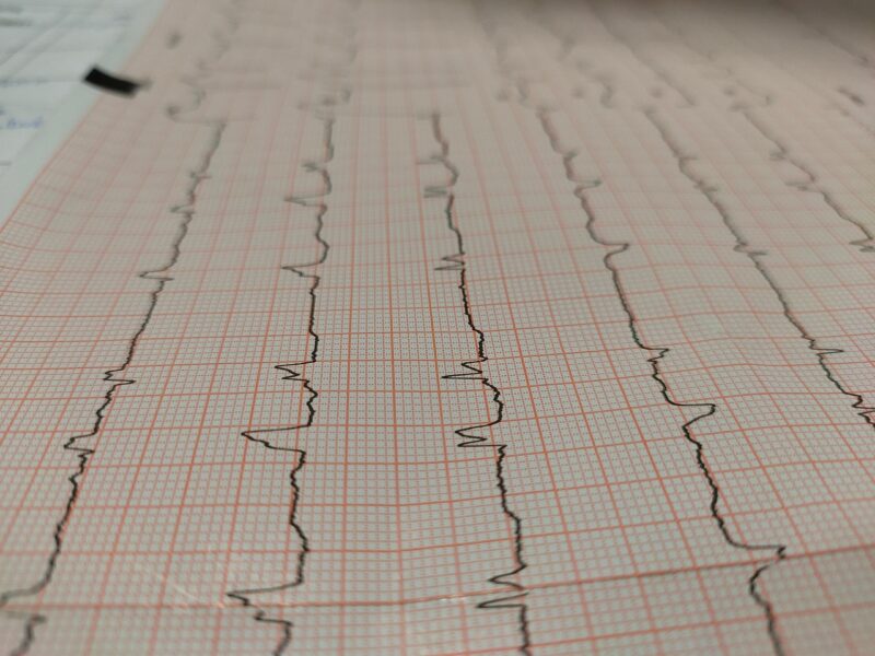
Electrocardiogram (ECG)
The standard 12-lead ECG remains the primary diagnostic tool for PVC identification. Medical literature indicates that ECGs can capture PVCs occurring during the brief recording period, though many PVCs may be missed due to their intermittent nature.
Ambulatory Monitoring
Extended monitoring provides more comprehensive assessment:
24-48 Hour Holter Monitoring: Studies show that continuous monitoring significantly increases PVC detection rates compared to standard ECGs.
Event Monitors: Research indicates that patient-activated monitors prove particularly useful for documenting symptomatic episodes.
Mobile Cardiac Telemetry: Advanced monitoring systems allow for extended observation periods with real-time analysis capabilities.
Echocardiography
Cardiac imaging studies help evaluate underlying structural heart disease that might predispose to PVCs or influence treatment decisions.
Management Strategies
Contemporary medical approach to PVC management emphasizes individualized assessment based on symptom severity, frequency, and underlying cardiac structure. Healthcare providers generally adopt a conservative approach for benign PVCs while addressing underlying causes.
Observation and Monitoring
Medical literature supports watchful waiting for asymptomatic individuals with infrequent PVCs and normal cardiac structure. Regular follow-up allows healthcare providers to monitor for changes in frequency or symptom development.
Lifestyle Modifications
Research consistently demonstrates the effectiveness of lifestyle interventions:
Trigger identification and avoidance: Studies suggest that identifying and minimizing personal PVC triggers can significantly reduce episode frequency.
Stress management techniques: Clinical trials indicate that stress reduction strategies may decrease PVC burden in susceptible individuals.
Sleep optimization: Sleep studies show that improving sleep quality and duration may reduce PVC occurrence.
Medical Intervention
Healthcare providers may consider pharmacological intervention in specific circumstances, though treatment decisions require careful individualization based on patient-specific factors and symptom severity.
Living with PVCs
Clinical experience demonstrates that many individuals with PVCs can maintain excellent quality of life through appropriate management and lifestyle adaptations. Understanding the generally benign nature of isolated PVCs helps reduce anxiety and improve coping strategies.
Quality of Life Considerations
Research indicates that patient education significantly impacts quality of life outcomes. Understanding that PVCs rarely represent serious cardiac pathology in structurally normal hearts often provides substantial reassurance.
Activity Modifications
Exercise physiology studies suggest that most individuals with benign PVCs can participate in regular physical activity. However, healthcare providers may recommend specific modifications based on individual circumstances.
Psychological Support
Medical literature recognizes the psychological impact of cardiac arrhythmias. Healthcare providers often recommend counseling or support groups for individuals experiencing significant anxiety related to PVC symptoms.
Prevention and Lifestyle Modifications
Preventive cardiology research identifies various strategies that may reduce PVC frequency and improve overall cardiac health. These evidence-based approaches focus on modifiable risk factors and healthy lifestyle promotion.
Dietary Considerations
Nutritional studies suggest several dietary modifications that may benefit individuals with PVCs:
- Maintaining adequate hydration
- Limiting excessive caffeine intake
- Ensuring proper electrolyte balance
- Following heart-healthy dietary patterns
Exercise and Physical Activity
Exercise physiology research demonstrates that regular, moderate physical activity generally benefits cardiac health, though individual responses may vary.
Stress Management
Behavioral medicine studies consistently show that effective stress management techniques can positively impact cardiac arrhythmia frequency.
FAQ
Q: Are PVCs dangerous? A: Medical research indicates that isolated PVCs in individuals with structurally normal hearts are generally benign. However, healthcare providers should evaluate concerning symptoms or frequent PVCs to rule out underlying conditions.
Q: Can PVCs be completely eliminated? A: Studies suggest that while PVC frequency can often be reduced through lifestyle modifications and medical management, complete elimination may not always be achievable or necessary.
Q: Do PVCs increase heart attack risk? A: Research shows that isolated PVCs in healthy individuals do not typically increase cardiovascular risk. However, underlying cardiac conditions that predispose to PVCs may carry independent cardiovascular risks.
Q: Can stress cause PVCs? A: Clinical observations consistently demonstrate associations between psychological stress and increased PVC frequency, likely through neuroendocrine mechanisms.
Q: Should I avoid exercise with PVCs? A: Exercise recommendations require individualized assessment by healthcare providers. Many individuals with benign PVCs can safely participate in regular physical activity.
Medical Disclaimer
This article provides educational information about premature ventricular contractions and should not replace professional medical advice, diagnosis, or treatment. Individual symptoms and circumstances vary significantly, requiring personalized healthcare provider evaluation. Always consult qualified medical professionals for specific concerns about cardiac symptoms or treatment decisions. Emergency medical attention should be sought immediately for chest pain, severe shortness of breath, or other concerning cardiac symptoms.
Sources
- Ataklte F, et al. Meta-analysis of ventricular premature complexes and their relation to cardiac mortality in general populations. Am J Cardiol. 2013;112(8):1263-70.
- Dukes JW, et al. Ventricular ectopy as a predictor of heart failure and death. J Am Coll Cardiol. 2015;66(2):101-9.
- Ng GA. Treating patients with ventricular ectopic beats. Heart. 2006;92(11):1707-12.
- Pedersen CT, et al. EHRA/HRS/APHRS expert consensus on ventricular arrhythmias. Heart Rhythm. 2014;11(10):e166-96.
- Yamada T, et al. Prevalence and clinical, electrocardiographic, and electrophysiologic characteristics of ventricular outflow tract tachycardia. Am J Cardiol. 1993;72(4):427-33.
This article is based on information from peer-reviewed medical literature and established clinical guidelines, including:
- American Heart Association Guidelines on Supraventricular Arrhythmias
- European Society of Cardiology Guidelines for Arrhythmia Management
- Journal of the American College of Cardiology publications on atrial tachycardia
- Heart Rhythm Society clinical recommendations
- New England Journal of Medicine cardiovascular research